How MRIs Can See Parkinson’s Coming Before Symptoms Appear


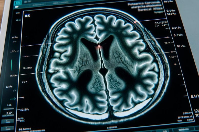
Researchers have found that MRI brain mapping can predict gray matter atrophy in mild Parkinson’s disease, offering a basis for personalized interventions to slow progression. Credit: SciTechDaily.com
New research reveals that MRI-based mapping of the brain’s structural and functional organization can predict the progression of gray matter atrophy in patients with early-stage, mild Parkinson’s disease.
The study utilized MRI data to generate a connectome, or brain map, to predict atrophy patterns, highlighting potential for tailored intervention trials and personalized treatment plans to combat disease progression.
Brain Mapping in Parkinson’s Disease
According to a study published today (June 25) in Radiology, the structural and functional organization of the brain as shown on MRI can predict the progression of brain atrophy in patients with early-stage, mild Parkinson’s disease. Radiology is a peer-reviewed scientific journal of the Radiological Society of North America (RSNA).
Parkinson’s disease is a progressive disorder characterized by tremors, slowness of movement or rigidity. Symptoms worsen over time and may include cognitive impairment and sleep problems. The disease affects more than 8.5 million people worldwide, and prevalence has doubled in the past 25 years, according to the World Health Organization (WHO).
Protein Clumps and Disease Mechanisms
One of the distinctive features of Parkinson’s disease is the presence of altered versions of the protein alpha-synuclein in the brain. Normally present in the brain, this protein accumulates as misfolded clumps inside nerve cells in Parkinson’s disease, forming structures known as Lewy bodies and Lewy neurites. These clumps spread to other brain regions, damaging nerves.
Researchers wanted to see if mapping the structural and functional connections across the brain could be used to predict patterns of atrophy spread in patients with mild Parkinson’s disease.
Study Design and Predictive Models
They used MRI data from 86 patients with mild Parkinson’s disease and 60 healthy control participants to generate the connectome, a structural/functional map of the brain’s neural connections. The researchers used the connectome to develop an index of disease exposure.
Disease exposure at one year and two years was correlated with atrophy at two years and three years post-baseline. Models including disease exposure predicted gray matter atrophy accumulation over three years in several areas of the brain.
“In the present study, brain connectome, both structural and functional, showed the potential to predict progression of gray matter alteration in patients with mild Parkinson’s disease,” said study coauthor Federica Agosta, M.D., Ph.D., associate professor of neurology at the Neuroimaging Research Unit of IRCCS San Raffaele Scientific Institute in Milan, Italy.
Potential Clinical Applications and Future Directions
The findings support the theory that functional and structural connections between brain regions may significantly contribute to Parkinson’s disease progression.
“The loss of neurons and accumulation of abnormal proteins can disrupt neural connections, compromising the transmission of neural signals and the integration of information across different brain regions,” Prof. Agosta said.
The study results point to a role for MRI in intervention trials to prevent or delay the disease progression—especially when individual patient information is incorporated into the model. Since Parkinson’s disease progression likely differs among individuals, future models should consider different starting conditions and incorporate individual-specific information for optimal effectiveness, according to Prof. Agosta.
“We believe that understanding the organization and dynamics of the human brain network is a pivotal goal in neuroscience, achievable through the study of the human connectome,” she said. “The idea that this approach could help identify different biomarkers capable of modulating Parkinson’s disease progression inspires our work.”
Reference: “Brain Connectivity Networks Constructed Using MRI for Predicting Patterns of Atrophy Progression in Parkinson’s Disease” 25 June 2024, Radiology.
Collaborating with Prof. Agosta were Silvia Basaia, Ph.D., Elisabetta Sarasso, M.Sc., Roberta Balestrino, M.D., Tanja Stojkovic, M.D., Ph.D., Iva Stanković, M.D., Ph.D., Aleksandra Tomić, M.D., Ph.D., Vladana Marković, M.D., Ph.D., Francesca Vignaroli, M.D., Elka Stefanova, M.D., Ph.D., Vladimir S. Kostić, M.D., Ph.D., and Massimo Filippi, M.D., F.E.A.N., F.A.A.N.


